What is Mohs Surgery? The Precise Skin Cancer Treatment
In the spirit of Melanoma Awareness Month, FoundHealth is disseminating information about the variety of integrative melanoma treatments and skin cancer treatments in general. In this week’s post, we are focusing on Mohs Micrograph Surgery, an extremely effective method for removing various skin cancers.
What is Mohs Surgery?
Background
Mohs surgery was actually invented in 1930′s, but has managed to withstand the test of time. Originally developed by Dr. Frederic E. Mohs, this method of skin cancer treatment became the choice method over the last decade after refinements were made. It is most effective in treating basal cell carcinoma and squamous cell carcinoma, the two most prevalent skin cancers. With regard to these two cancers, Mohs Surgery touts a success rate between 96-98%, depending on the data source. In the past, Mohs method was rarely used for treating melanoma (the most dangerous of the skin cancers), for fear that the microscopic melanoma cells would be missed and would then metastasize(spread) throughout the body. However, highly specialized stains have recently been developed that allow melanoma cells to be more easily visualized, and this breakthrough has caused Mohs surgery to be used more frequently for treatment of melanoma.
The Method – Step by Step
Mohs surgery is a unique skin cancer treatment because it maximizes the chance of removing all the cancerous cells while minimizing the amount of skin that is removed.
 Step 1- As shown in the image, the visible portion of the skin cancer often does not indicate the full extent of the cancer. Instead, the cancer is rooted deeper into the skin. Mohs surgery is unique because rather than making an educated guess as to where the cancer may stop, the skin samples are analyzed during the surgery, and all the identified carcinoma roots are removed.
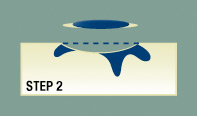
 Step 2- The surgeon will remove the visible cancer along with 1 to 1.5 mm of surrounding, healthy looking skin. The amount of healthy looking skin removed is known as the ‘surgical margin’. In most skin cancer treatments, an average size of the surgical margin is 4-6 mm.
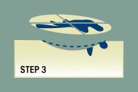
 Step 3- Now that the visible tumor has been removed, a thin layer of underlying skin is removed using a sharp, precise scalpel. Using a cryostat (a device used to keep the skin sample cool) and various dyes, the Mohs surgeon sections the skin sample and makes a map that shows how the skin sample corresponds to the skin removal site.
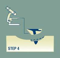
Step 4- The edges of the skin sample are examined under a microscope, and evidence of cancerous cells are noted on the map. Once the sample has been 100% examined, the Mohs surgeon returns to the patient and, using the map, he removes additional layers of skin from the regions where evidence of cancerous cell roots exist.
 Step 5- The doctor will examine these new sections of removed skin, and perform the same steps of analysis. If he finds additional cancer cells on the margins of the samples, he will remove more skin. If he does not, it is an indication that all the cancer is removed, and the procedure is concluded.
If you have an experience with Mohs surgery, please share your feedback with the community!
Join Our Community
Archives
- January 2023
- December 2022
- September 2022
- August 2022
- June 2022
- May 2022
- April 2022
- March 2022
- February 2022
- January 2022
- December 2021
- November 2021
- October 2021
- September 2021
- August 2021
- July 2021
- June 2021
- May 2021
- March 2021
- September 2020
- August 2020
- July 2020
- June 2020
- May 2020
- April 2020
- March 2020
- February 2020
Subscribe

Sign up to receive FREE toolkit
From Dr. Hyman, #1 NY Times & Amazon Author
We never spam or sell your e-mail


Follow Our Every Move